Introduction
This Atlas was developed to identify the normal neuroanatomical structures of the entire brain in the MRI scan. This author hopes that the Atlas would help:- Teaching, testing, and learning the brain’s anatomy through interactive technology.
- Correlating accurately the location of lesions found on patients’ MRI scans with the anatomical structures.
- To know the topographic distances of anatomic structures from the cortex and central baselines during neurosurgical operations.
During intracranial operations, as one attempts to reach a lesion or remove an infiltrating one, knowing the location becomes essential as one operates deeper; the three-dimensional measured distances in this Atlas should help.
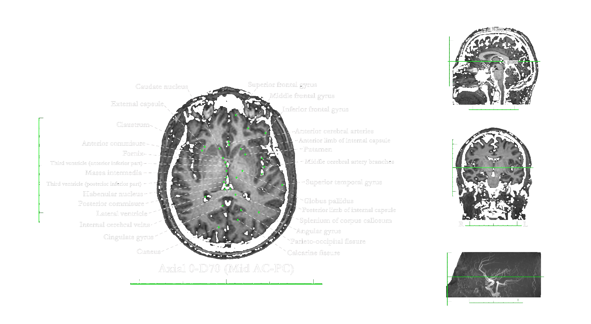
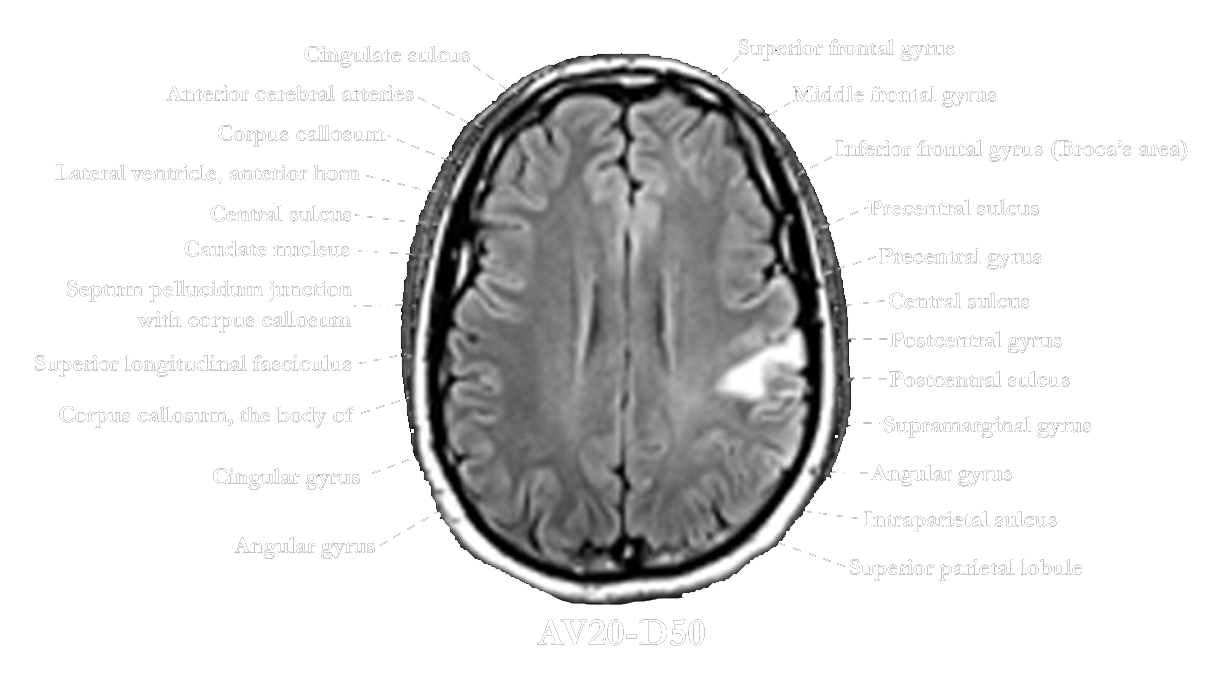
The MRI scan is that of a 31-year-old healthy male with no history of any abnormal neurological condition. It is one of the 10 MRI scans of healthy individuals, obtained with IRB approval to study the anatomical correlation of the brain with the MRI scan, taken in a 3Tesla scanner. This case was selected because more views were available.
The atlas is uploaded to the Internet. Free, only for educational purposes. It shows best in computers. It also shows on tablets and cell phones. However, the terms are small on cell phones.
Professor of Neurological Surgery
The University of Chicago

This work is licensed under a Creative Commons Attribution-NonCommercial-NoDerivatives 4.0 International License.