How To Use The Interactive Interface
Abbreviations
|
A = Axial
B = Basal C = Coronal D = Distance from surface of cortex F = Frontal
O = Occipital V = Vertex L = Left R = Right AC = Anterior Commissure
PC = Posterior Commissure Mid AC-PC = A point in the middle of AC-PC line, 0 point |
Topographic Divisions

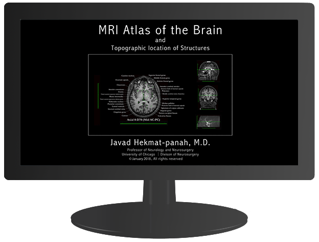
Mid AC-PC point: A point just in the middle of the AC-PC line. This point can be considered as 0 point where axial, coronal, and sagittal planes cross.
Axial Plane: is at the level of AC-PC line, dividing the brain into:
Axial Vertex (AV) and Axial Base (AB)
Basal Plane: is parallel to Axial Plane, separating the Axial Base (AB) from spinal cord.
Coronal Plane: is perpendicular to the axial plane and goes through the mid AC-PC point, dividing the brain into:
Coronal Frontal (CF) and Coronal Occipital (CO)
Sagittal plane: is between the two hemispheres, dividing the brain into:
Sagittal Left (SL) and Sagittal Right (SR)
Brain Sections: each MRI section is 5mm apart, starting from axial, coronal, and sagittal planes.
Sections' Name: are based on their topographic location from their corresponding planes. Each section is hyphenated with two names. The first half of the name determines its distance from the corresponding plane. The second half is its distance (D) from the most outer surface of cerebral cortex of that plane; or in Axial Base Sections, from the basal plane.
The second half is included to conceptually help localization of a lesion on MRI scans of the brain or when one is working in supine, lateral, or prone positions during intracranial operations.
The additional smaller views help orient the user in the 3-dimensionality of each section.
Examples
The Topographic Divisions of the brain are defined according to their relation to the AC-PC line, a line drawn from the upper surface of the Anterior Commissure (AC) horizontally to the Posterior Commissure (PC) in sagittal view.The following examples help indicate what the abbreviations represent.
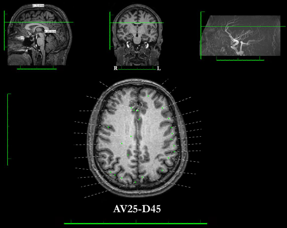
In Axial Vertex Sections: AV25-D45 is the abbreviation of Axial Vertex: 25mm from the axial plane, with a Distance of 45mm from the vertex cortex.
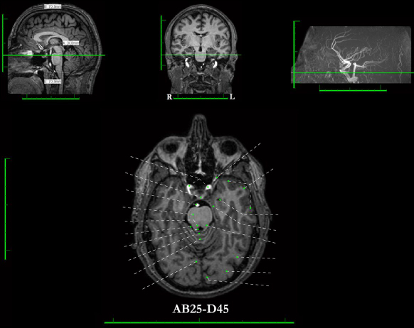
In Axial Base sections: AB25-D45 is the abbreviation of Axial Base: 25mm from the axial plane, with a Distance of 45mm from Basal plane.
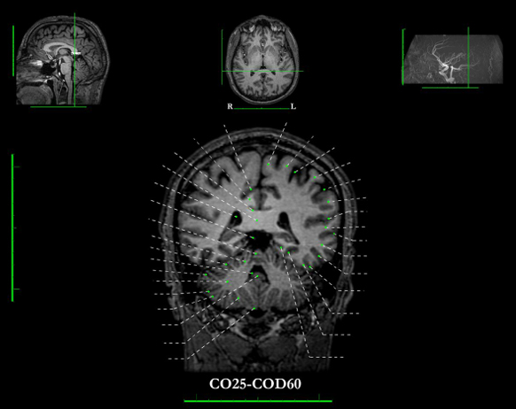
In Coronal Section: CO25-COD60 is the abbreviation of Coronal Occipital: 25mm from the coronal plane, with a Distance of 60mm from the most posterior end of occipital cortex.
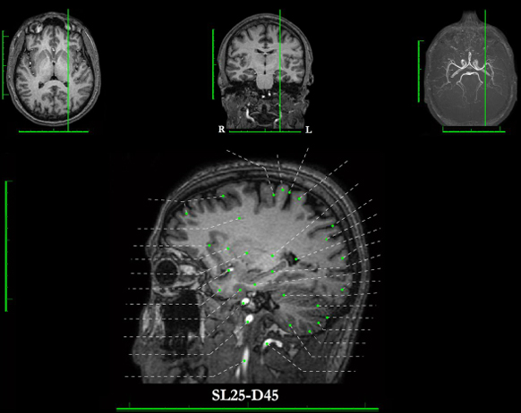
In Sagittal Sections: SL25-D45 is the abbreviation of Sagittal Left: 25mm left of the sagittal plane, with a Distance of 45mm from the most lateral extent of the left cortex.
JHP MRI Atlas of the Brain

This work is licensed under a Creative Commons Attribution-NonCommercial-NoDerivatives 4.0 International License.
• The author appreciates the Brain Research Foundation’s support in obtaining the MRI scans.
• The University of Chicago Research Computing Center provided technical support, uploading, and hosting this atlas. The author appreciates the center's continued support in maintaining and the need for periodic rearrangements.
• Henry Gray and Susan Standring, Gray's Anatomy. Elsevier Academic Press, 2008.
• Zang-Hee Cho, editor, 7.0 Tesla MRI Brain Atlas. Springer, 2010.
• Melville P. Roberts et al., Atlas of the Human Brain in Section.
Lippincott Williams & Wilkins, 1987.
• "Salamonís Neuroanatomy and Neurovasculature Web-Atlas Resource"
http://www.radnet.ucla.edu/sections/DINR
• Jurgen K. Mai et al., Atlas of the Human Brain, second edition.
Elsevier Academic Press, 2004.